Pleural Mesothelioma X Ray : Mesothelioma Pulmonary Disorders Msd Manual Professional Edition - None of the scans cause pain for the patient.. How does mesothelioma affect lung capacity? Unfortunately, there is no one single tool that can always confirm or rule out mesothelioma, and many patients have to undergo multiple tests with each test having the possibility of pointing to another condition like pneumonia first. However, when a tumor is present on or near the lungs (such as on the pleural lining), doctors may see an opaque white area. Treating mesothelioma vs pleural plaques Chest x ray is the initial screening test for the mesothelioma like all other the chest diseases.
Chest x ray is the initial screening test for the mesothelioma like all other the chest diseases. No evidence of primary tumor; Primary tumor cannot be assessed; Treating mesothelioma vs pleural plaques Doctors will determine a pleural mesothelioma diagnosis by ordering:
Https Encrypted Tbn0 Gstatic Com Images Q Tbn And9gcs5ch5a1ipmcair5cf2qg7wafbstxn0ezkqpfrhnwvwovtro3ic Usqp Cau from Below is the eighth edition of the tnm staging system for malignant pleural mesothelioma, which was published in 2018 1. How does mesothelioma affect lung capacity? More specialized imaging scans and tissue biopsies to confirm a positive diagnosis for pleural mesothelioma, including: Involving ipsilateral parietal pleura (inc. Mesothelioma isn't always fatal and that's the hope to hold on to. If imaging tests show signs of cancer, biopsies are ordered to extract tissue or fluid through a needle to test for cancer cells. Diagnosing pleural mesothelioma often consists of multiple tests. People diagnosed with mesothelioma have aggressive cancer that is caused by asbestos exposure.
Tumors on or around the lungs;
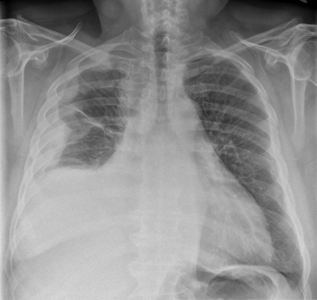
None of the scans cause pain for the patient. But computed tomography (ct) is the imaging technique of choice for charactering pleural masses. More specialized imaging scans and tissue biopsies to confirm a positive diagnosis for pleural mesothelioma, including: This position is called lateral decubitus position. Chest x ray is the initial screening test for the mesothelioma like all other the chest diseases. This excess fluid is called pleural effusion, a common symptom of pleural mesothelioma. Primary tumor cannot be assessed; Pleural mesothelioma is caused by inhaling asbestos fibers that get lodged into the protective lining of the lungs (the pleura) which cause genetic mutations in the surrounding cells. Ct scans and mris for mesothelioma diagnosis Given the presence of the mesothelium in different parts of the body, mesothelioma can arise in various locations 17:. However, when a tumor is present on or near the lungs (such as on the pleural lining), doctors may see an opaque white area. Ct scans for mesothelioma we have all heard of ct or cat scans. The body naturally produces small amounts of pleural fluid in order to lubricate the surfaces of the pleura, which is a serous membrane located within the pleural cavity that surrounds the lungs.
One manner in which malignant pleural mesothelioma can affect a patient's lung capacity is through a process known as pleural effusion. Mesothelioma x ray, asbestosdefinition.com | mesothelioma primarily is composed of 3 distinct types. The length of the process varies for each type of imaging test. More specialized imaging scans and tissue biopsies to confirm a positive diagnosis for pleural mesothelioma, including: Find updated content daily for what mesothelioma
Mesothelioma Radiology Case Radiopaedia Org from prod-images-static.radiopaedia.org However, the presence of a pleural effusion could also indicate other abnormalities in the lung or the pleura. For example, it can relieve symptoms such as pain caused by tumours, and it can be given after chemotherapy or surgery, to help kill remaining cancer cells. Ct scans for mesothelioma we have all heard of ct or cat scans. Chest x ray is the initial screening test for the mesothelioma like all other the chest diseases. Unfortunately, there is no one single tool that can always confirm or rule out mesothelioma, and many patients have to undergo multiple tests with each test having the possibility of pointing to another condition like pneumonia first. Tumors on or around the lungs; The length of the process varies for each type of imaging test. Imaging plays an essential role in the diagnosis, staging, and clinical management of patients with mesothelioma x ray imaging techniques chest radiography and computed tomography (ct), magnetic resonance imaging (mri), and positron emission tomography (pet) have all been used to evaluate this disease, although the relative importance of these imaging modalities has changed over time.
The length of the process varies for each type of imaging test.
Ct scans and mris for mesothelioma diagnosis The length of the process varies for each type of imaging test. Mesothelioma x ray, asbestosdefinition.com | mesothelioma primarily is composed of 3 distinct types. Most tumors arise from the pleura, and so this article will focus on pleural mesothelioma. Unfortunately, there is no one single tool that can always confirm or rule out mesothelioma, and many patients have to undergo multiple tests with each test having the possibility of pointing to another condition like pneumonia first. For example, it can relieve symptoms such as pain caused by tumours, and it can be given after chemotherapy or surgery, to help kill remaining cancer cells. Some are better at detecting pleural mesothelioma than peritoneal mesothelioma. Imaging plays an essential role in the diagnosis, staging, and clinical management of patients with mesothelioma x ray imaging techniques chest radiography and computed tomography (ct), magnetic resonance imaging (mri), and positron emission tomography (pet) have all been used to evaluate this disease, although the relative importance of these imaging modalities has changed over time. Pleural mesothelioma makes up approximately 75 percent of all mesothelioma cases, meaning specialists have more opportunities to develop new treatments each year. Doctors will determine a pleural mesothelioma diagnosis by ordering: Find updated content daily for what mesothelioma However, when a tumor is present on or near the lungs (such as on the pleural lining), doctors may see an opaque white area. One manner in which malignant pleural mesothelioma can affect a patient's lung capacity is through a process known as pleural effusion.
Unfortunately, there is no one single tool that can always confirm or rule out mesothelioma, and many patients have to undergo multiple tests with each test having the possibility of pointing to another condition like pneumonia first. For example, it can relieve symptoms such as pain caused by tumours, and it can be given after chemotherapy or surgery, to help kill remaining cancer cells. Pleural mesothelioma makes up approximately 75 percent of all mesothelioma cases, meaning specialists have more opportunities to develop new treatments each year. Treating mesothelioma vs pleural plaques Find updated content daily for what mesothelioma
Malignant Mesothelioma Radiographics from pubs.rsna.org Tried, tested, trusted and affordable for all qpcr needs. Mesothelioma is a rare form of cancer typically affecting the lining of the lungs. One manner in which malignant pleural mesothelioma can affect a patient's lung capacity is through a process known as pleural effusion. Pleural mesothelioma makes up approximately 75 percent of all mesothelioma cases, meaning specialists have more opportunities to develop new treatments each year. Doctors may also use one or more other imaging tests to make a diagnosis. None of the scans cause pain for the patient. Involving ipsilateral parietal pleura (inc. Ct scans for mesothelioma we have all heard of ct or cat scans.
Pleural mesothelioma is caused by inhaling asbestos fibers that get lodged into the protective lining of the lungs (the pleura) which cause genetic mutations in the surrounding cells.
Below is the eighth edition of the tnm staging system for malignant pleural mesothelioma, which was published in 2018 1. Mesothelioma awareness and understanding its signs is especially important for helping to diagnose mesothelioma at an early stage. Ct scans and mris for mesothelioma diagnosis Pleural mesothelioma makes up approximately 75 percent of all mesothelioma cases, meaning specialists have more opportunities to develop new treatments each year. It is very important to realize that imaging tests alone can't diagnose mesothelioma. Ct also gives important information regarding invasion of the chest wall and surrounding structures. It's often difficult to diagnose mesothelioma by viewing cells from fluid samples. One manner in which malignant pleural mesothelioma can affect a patient's lung capacity is through a process known as pleural effusion. Pleural mesothelioma is caused by inhaling asbestos fibers that get lodged into the protective lining of the lungs (the pleura) which cause genetic mutations in the surrounding cells. The body naturally produces small amounts of pleural fluid in order to lubricate the surfaces of the pleura, which is a serous membrane located within the pleural cavity that surrounds the lungs. They can schedule additional tests to learn more about what is causing symptoms. However, the presence of a pleural effusion could also indicate other abnormalities in the lung or the pleura. Treating mesothelioma vs pleural plaques
Ct also gives important information regarding invasion of the chest wall and surrounding structures mesothelioma x ray. Diagnosing pleural mesothelioma often consists of multiple tests.
0 Response to "Pleural Mesothelioma X Ray : Mesothelioma Pulmonary Disorders Msd Manual Professional Edition - None of the scans cause pain for the patient."


0 Response to "Pleural Mesothelioma X Ray : Mesothelioma Pulmonary Disorders Msd Manual Professional Edition - None of the scans cause pain for the patient."
Post a Comment